Mass Spectrometry Core
 Mass Spectrometry Core
Van Andel Institute’s Mass Spectrometry Core is committed to developing scientific partnerships with research labs to develop and deploy LC/MS and GC/MS methods specifically tailored to meet the needs of every project. The Core houses a comprehensive suite of technologies and staff expertise to deliver high-quality metabolomics, lipidomics and proteomics capabilities to VAI scientists and collaborators.
For questions, please contact Core Director Dr. Ryan Sheldon.
Recent News & Publications
Learn More
Cancer cells can use backup routes to fuel their growth

‘Sweet’ discovery reveals how glucose fuels cancer-fighting immune cells

Study finds persistent proteins may influence metabolomics results
Madaj ZB, Dahabieh MS, Kamalumpundi V, Muhire B, Pettinga J, Siwicki RA, Ellis AE, Isaguirre C, Escobar Galvis ML, DeCamp L, Jones RG, Givan SA, Adams M, Sheldon RD. 2023. Prior metabolite extraction fully preserves RNAseq quality and enables integrative multi-omics analysis of liver metabolic response to viral infection. RNA Biol 20(1).
Sheldon RD, Ma EH, DeCamp LM, Williams KS, Jones RG. 2021. Interrogating in vivo T-cell metabolism in mice using stable isotope labeling metabolomic and rapid cell sorting. Nat Proto.
Izreig S, Gariepy A, Kaymak I, Bridges HR, Donayo AO, Bridon G, DeCamp LM, Kitchen-Goosen SM, Avizonis D, Sheldon RD, Laister R, Minden MD, Johnson NA, Duchaine TF, Rudoltz MS, Yoo S, Pollak MN, Williams KS, Jones RG. 2020. Repression of LKB1 by miR-17~92 sensitizes MYC-dependent lymphoma to biguanide treatment. Cell Rep Med.
Our Impact
We’re raising thousands to save millions.
We’re turning hope into action for the millions of people around the world affected by diseases like cancer and Parkinson’s. Find out how you can help us make a difference.
- 122 peer-reviewed papers published in 2024, 63 of which were in high-impact journals
- 15 VAI-SU2C Epigenetics Dream Team clinical trials launched to date
- 10 clinical trials co-funded by VAI & Cure Parkinson's (out of 41 total International Linked Clinical Trials Program trials)
Metabolomics and Lipidomics
Mass Spectrometry Core employs mass spectrometry-chromatography based approaches for targeted and untargeted metabolite profiling, stable isotope tracing to understand metabolite movement, lipidomics analysis, and absolute metabolite quantitation.
The metabolome consists of hundreds to thousands of small molecules (< 50 Da to > 2000 Da) with vastly different chemistries; no single method is suitable to assess it in its entirety. To account for this, the Mass Spectrometry Core offers a range of chromatographic and MS methods to best address each research question.
- Targeted metabolomics experiments are used to identify compounds in a pre-selected metabolite class or pathway. Targeted metabolite panels have been developed to provide high-confidence compound identification through direct comparison to known chemical standards.
- Untargeted metabolomics analysis identifies all features in an experimental run and uses library matching for compound identification.
A metabolic tracing experiment provides an understanding of the metabolic flux within a biological model. In these studies, the researcher introduces a heavy stable isotope, such as 13C, into the experimental system. Then, the mass spectrometer detects alterations in the isotope patterns to determine the fraction of each metabolite pool that contains the heavy atom(s). These analyses can be either targeted or untargeted.
The Mass Spectrometry Core can provide guidance for the experimental design and sample handling for tracing experiments. For each experiment, the researcher must provide unlabeled control samples to be used for correction for naturally occurring isotopes already present in the system.
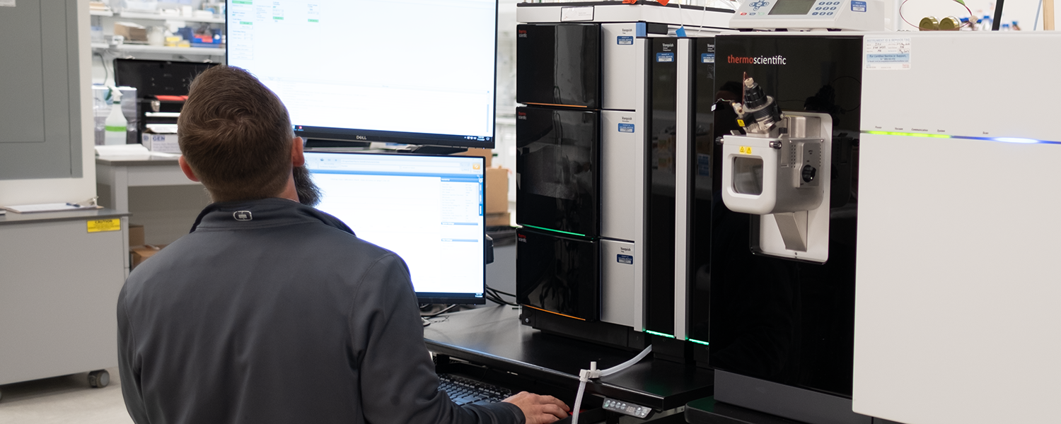
The Core has developed a lipidomics platform using the Thermo Scientific Orbitrap ID-X LC/MS, which can routinely annotate 500+ lipid species including phospholipids, ceramides, triacylglycerides, cardiolipins and cholesterol esters. The lipidomics method is semi-targeted, relying on previously run standards to confidently identify features, while also detecting any other features present in the sample.
For increased confidence in lipid identification, the EquiSPLASH®, Avanti Polar Lipids standard mixture can be used. The mixture of 13 deuterated lipid internal standards will be added to all samples, quality controls and blanks at a final resuspended concentration of 25 ug/mL post-metabolite extraction. This control serves to validate the accuracy of acquired data and, if needed, help normalize data for any fluctuations in metabolite abundances that are not the result of biological differences between samples. Due to the cost of the standard mixture, an additional fee is required for this option.
The metabolomics services described above provide relative quantitation, allowing comparison of metabolite pool sizes across experimental groups within a metabolite. Absolute quantitation requires preparation and analysis of the target metabolite(s) at known concentrations (i.e., a dilution curve of the chemical standard). The response of each known concentration can be used to quantitate the response from biological samples through a regression analysis.
An additional control required for targeted quantitation experiments is an isotopically labeled internal standard added to both the experimental and quantitation samples. The internal standard is used to correct for matrix effects as well as expected instrument variability during MS analysis.
Targeted quantitation experiments require metabolite- and matrix-specific method development to ensure accurate quantitation. The MSC routinely develops such methods, but planning, time, and researcher-provided pilot samples are required. The researcher must also provide the neat chemical standard(s) and isotopically labeled internal standard(s) to be used. After the initial method development, the method can be used routinely. The quantitation samples and internal standard will be run with all subsequent experiments.
Proteomics
The Core employs a variety of mass spectrometry-based experiments to characterize and quantify proteins from complex biological samples. We offer a range of services, including global protein/PTM quantitation, interactome analysis, PTM characterization, targeted assays, as well as advanced statistical and bioinformatics analysis.
During the past two decades, mass spectrometry (MS) has been established as the primary method for protein identification from complex mixtures of biological origin.
Immunoprecipitation (IP)/pull-down experiments leveraging MS provide sensitive and accurate characterization of protein complexes. Characterizing proteins of interest using this workflow is one of our most popular experiments. In these studies, a protein complex to be analyzed is first purified using an appropriate approach. Isolated proteins are proteolytically digested (most often using trypsin) to generate a mixture of peptides that can be identified by MS.
- Protein and post-translational modification (PTM) characterization. Mass spectrometry analysis after immunoprecipitation is a powerful approach for characterizing proteins of interest and their posttranslational modifications (PTMs).
- Profiling the partners of a given protein (Interactome). Protein-protein interactions and components of multiprotein complexes can be characterized.
- Protein profiling from a complex mixture. Identifying and characterizing the different types of proteins present in a complex sample.
High-resolution mass spectrometry enables the quantification of hundreds to thousands of proteins across different biological conditions. This powerful analysis can provide deep insights into biology’s fundamental mechanisms and processes by measuring differential protein expression.
In the past, mechanistic investigations have traditionally focused on one or a few proteins. However, advances in MS technology have made it possible to examine large networks of molecules in a single study. Currently available MS technologies can identify 10 peptides per second with ppm mass accuracy and enables the detection of more than 5,000 proteins in two hours (depending on sample type). Our global proteome quantification method attempts to accurately identify and quantify as many proteins as possible in a sample.
- Tandem Mass Tag (TMT): Isobaric chemical tags represent a fast, unbiased and sensitive method to quantify proteins in biological samples. Tandem Mass Tag (TMT, Thermo Fisher Scientific) are isobaric chemical tags that provide multiplexing capabilities for determining the relative abundance of proteins in proteomics experiments. The ability to perform concurrent MS analysis on multiple samples increases throughput and enables relative quantitation of up to 16 different samples derived from cells, tissues, or biological fluids.
- Data-Independent Acquisition (DIA): Data-independent acquisition (DIA) has emerged as an alternative to comprehensive identification and precise quantification. DIA carries out fragmentation using wider isolation widths (typically 10–50 Da), covering all m/z values within a certain mass range. By fragmenting all ions within this mass range, more signals will be generated for a single peptide, giving a more reliable relative quantification than conventional label-free approaches.
We strongly suggest conducting a DIA experiment for global proteomics, except in cases where the experiments are solely suited for the TMT platform.
Targeted assays enable the detection and quantification of a predetermined subset of proteins with high sensitivity and reproducibility across many samples. Additionally, targeted analyses can measure the relative abundance of hundreds of proteins simultaneously and accurately. For this assay, synthetic light or heavy isotope-labeled peptides of the target proteins may be required.
- Parallel-reaction monitoring (PRM): This assay provides high selectivity, high sensitivity and high-throughput relative quantification with confident targeted peptide confirmation.
Bioinformatics Expertise
The Mass Spectrometry Core has expertise in software and informatics pipelines for conducting metabolomics and proteomics investigations. Computational services include data processing, imputation, statistical analyses, data visualization and various specialized bioinformatics analyses. The Core also supports custom computational analyses for metabolomics or proteomics studies through the VAI Bioinformatics and Biostatistics Core.
Metabolomics and Lipidomics
Three liquid chromatography-mass spectrometry (LC/MS) systems and three gas chromatography (GC)/MS systems are dedicated to small molecule analysis. These instruments deploy an array of standard methods, annotated with a chemical library of more than 1,000 compounds, to maximize compound coverage in every sample.
For metabolomics, these methods routinely detect 200+ compounds including TCA cycle intermediates, glycolysis, nucleotides, amino acids, urea cycle, vitamins, cofactors tryptophan/kynurenine metabolism, acyl-CoA’s, acyl-carnitines and more. For lipidomics, 500+ lipid species are routinely annotated including phospholipids, ceramides, triacylglycerides, cardiolipins, cholesterol esters, and more. These methods can be operated in an untargeted manner, allowing for screening for biomarkers and novel metabolites/lipids, and in the context of stable isotope tracing, for assessment of metabolic movement. Further, upholding the scientific enterprising mission of the Mass Spectrometry Core, our team routinely develops custom methods to quantitate project-specific compounds of interest.
Primary use: Semi-untargeted metabolite and lipid profiling and stable isotope labeling
Capabilities
- High-resolution/accurate mass data acquisition
- Up to four chromatographic separations per sample to maximize metabolite coverage
- MS2 fragmentation for compound identification and library matching
- MSn fragmentation for structural elucidation
Specifications
- Up to 500K resolution (FWHM)
- < 2 ppm mass accuracy
- Electrospray ionization
- Dual Vanquish UHPLC
- Data dependent acquisition
- CID and HCD fragmentation
Primary use: Stable isotope labeling of central carbon metabolism
Capabilities
- High-resolution/accurate mass data acquisition
- Duplexed ion-paired chromatography for reproducible retention times and maximum throughput
- MS2 fragmentation for compound identification and library matching
Specifications
- 240K resolution (FWHM)
- < 2 ppm mass accuracy
- Electrospray ionization
- Data-dependent acquisition
Primary use: Targeted polar metabolite profiling and custom targeted method development
Capabilities
- Ion-paired negative mode profiling of ~250 polar metabolites, including amino acids, nucleotides, carboxylates and sugar phosphates
- dMRM mode limits transition monitoring only to retention windows of interest to maximize compounds detected per run
- Highly reproducible ion-paired chromatography
- High sensitivity and specificity of targeted compounds
- Second UHPLC system for targeted method development of compounds not amenable to ion-pairing.
Specifications
- Sub-femtogram detection limits (compound dependent)
- Electrospray ionization
Primary use: Targeted metabolite profiling of central carbon metabolism and custom targeted method development
Capabilities
- Profiling, quantitation and stable isotope labeling of most amino acids and TCA cycle intermediates
- In-source compound fragmentation for positive ID
Specifications
- Sub-femtogram detection limits (compound dependent)
- Electron-impact ionization
The rates of oxygen consumption (OCR; i.e., mitochondrial ATP production) and extracellular acidification (ECAR; proportional to glycolytic rate in most systems) in live cells are determined in a convenient 96-well format under a variety of stimulation conditions. These data are often most robust when combined with metabolomic approaches as described above. The Seahorse can give functional significance to metabolomics findings; for example, if glucose more strongly labels the TCA cycle than glutamine in a SIL study, then this can be confirmed by OCR in the presence of glucose or glutamine. Importantly, a lack of phenotypic differences by Seahorse does not preclude a metabolomic phenotype.
Primary use: Measure OCR and ECAR of live cells in a 96-well plate format. OCR and ECAR rates are key indicators of mitochondrial respiration and glycolysis; these measurements provide a systems-level view of cellular metabolic function in cultured cells and ex vivo samples
Capabilities
- Four-port injection system and automated mixing allows real-time detection of responses to substrates, inhibitors and other compounds. Temperature is maintained at 37o
- A 96-well plate allows multiple conditions to be run at once. The system is optimized for compound screening and dose-response studies.
- As few as 5,000 cells per well may be analyzed at high sensitivity (depending on cell type)
- System is compatible with many different sample types
Specifications
- 96 assay wells
- 2 µL microchamber volume
Proteomics
In 2021, the Core expanded its capabilities to include proteomics with the addition of a Thermo Orbitrap Eclipse mass spectrometer. This equipment provides investigators with access to the most advanced high-resolution MS-based proteomics technologies. We employ a variety of mass spectrometry-based experiments to identify, quantify and characterize proteins from complex biological samples. We anticipate adding another dedicated proteomics LC/MS system in 2023, and additional proteomics instrumentation as needed over the next five years.
Primary use: Bottom-up and middle-down proteomics applications
Capabilities
- High-resolution/accurate mass data acquisition
- MSn fragmentation
- Data-dependent acquisition and data-independent acquisition
- Real-time search for TMT quantification
- High-sensitivity nano-flow UPLC
Specifications
- Up to 500K resolution (FWHM)
- < 2ppm mass accuracy
- Electrospray ionization
- CID, HCD and ETD fragmentation
- High field asymmetric waveform ion mobility (FAIMS)
- High mass range

Ryan Sheldon, Ph.D.
Core Director, Mass Spectrometry Core

Colt Capan, M.S.
Core Associate, Mass Spectrometry Core

Abigail Ellis, B.S.
Core Laboratory Manager, Mass Spectrometry Core

Megan Gendjar, M.S.
Core Associate, Mass Spectrometry Core

Molly Hopper, Ph.D.
Core Scientist, Mass Spectrometry Core

Christine Isaguirre, B.S.
Core Project Manager, Mass Spectrometry Core

Amy Johnson, B.S.
Core Associate, Mass Spectrometry Core

Hyoungjoo Lee, Ph.D.
Core Scientist, Mass Spectrometry Core

Emilie Poirier
Student Intern, Mass Spectrometry Core

Ibukunoluwa Sodiya, M.S.
Core Associate, Mass Spectrometry Core

Anna Tarach, B.S.
Core Associate, Mass Spectrometry Core

Michael Vincent, Ph.D.
Core Bioinformatics Scientist, Mass Spectrometry Core
Acknowledgments and Authorship
All work performed by VAI’s Core Technologies and Services should be acknowledged or considered for co-authorship in scholarly reports, presentations, posters, papers and all other publications. Proper acknowledgment allows us to obtain financial and other support that enables us to provide and maintain high-quality support and services. By acknowledging shared resource facilities and instrumentation in publications, presentations and other research communications, you play a critical role in ensuring their continued availability and future development.
Example acknowledgments:
- We thank the Van Andel Institute Mass Spectrometry Core (RRID:SCR_024903), especially [staff name], for their assistance with [technique/technology].
- This research was supported in part by the Van Andel Institute Mass Spectrometry Core (Grand Rapids, MI) (RRID:SCR_024903).
PUBLICATIONS
Please note, the Mass Spectrometry Core opened in fall 2019. Papers will be listed here as they are published.
Nauta KM, Gates DR, Weiland M, Mechan-Llontop ME, Wang X, Isaguirre C, Gendjar MR, Cooper J, Barton SF, Nguyen KP, Sheldon RD, Krawczyk CM, Burton NO. 2025. A noncanonical polyamine from bacteria antagonizes host mitochondrial function. Nat Commun.
Forton C, DeVries J, Lou M, Brundin S, Cave T, Anis E, Madaj ZB, Isaguirre C, Johnson A, Sheldon RD, Smart L, Bohnert KM, Kassien J, Holzgen O, Youssef NA, Khan T, Brundin L. 2025. Gut microbiome‑derived tryptophan metabolites predict relapse in alcohol use disorder. Brain Behav Immun.
Kasen A, Lövestam S, Breton L, Meyerdirk L, McPhail JA, Piche K, Louwrier A, Capan CD, Lee H, Scheres SH, Henderson MX. 2025. Seed structure and phosphorylation in the fuzzy coat impact tau seeding competency. Nat Commun 16(1).
Longo J, Watson MJ, Williams KS, Sheldon RD, Jones RG. 2025. Nutrient allocation fuels T cell-mediated immunity. Cell Metab.
Kaluba FC, Rogers TJ, Jeong YJ, House RJ, Waldhart A, Sokol KH, Daniels SR, Lee CJ, Longo J, Johnson A, Sartori V, Sheldon RD, Jones RG, Lien EC. 2025. An alternative route for β-hydroxybutyrate metabolism supports cytosolic acetyl-CoA synthesis in cancer cells. Nat Metab.
Pérez-Mojica JE, Madaj ZB, Isaguirre C, Roy J, Lau KH, Sheldon RD, Lempradl A. 2025. Resolving early embryonic metabolism in Drosophila through single-embryo metabolomics and transcriptomics. Nat Metab.
*Featured on the cover
Longo J, DeCamp LM, Oswald BM, Teis R, Reyes-Oliveras A, Dahabieh MS, Ellis AE, Vincent MP, Damico H, Gallik KL, Foy NM, Compton SE, Capan CD, Williams KS, Esquibel CR, Madaj ZB, Lee H, Roy DG, Krawczyk CM, Haab BB, Sheldon RD, Jones RG. 2025. Glucose-dependent glycosphingolipid biosynthesis fuels CD8+ T cell function and tumor control. Cell Metab.
Dahabieh MS, DeCamp LM, Oswald BM, Kitchen-Goosen SM, Fu Z, Vos M, Compton SE, Longo J, Foy NM, Williams KS, Ellis AE, Johnson A, Sodiya I, Vincent M, Lee H, Yao C, Wu T, Sheldon RD, Krawczyk CM, Jones RG. 2025. The prostacyclin receptor PTGIR is a NRF2-dependent regulator of CD8+ T cell exhaustion. Nat Immunol.
*Featured in a News & Views article
Kaymak I, Watson MJ, Oswald BM, Ma S, Johnson BK, DeCamp LM, Mabvakure BM, Luda KM, Ma EH, Lau KH, Fu Z, Muhire B, Kitchen-Goosen SM, VanderArk A, Dahabieh MS, Samborska B, Vos M, Shen H, Fan ZP, Roddy TP, Kingsbury GA, Sousa CM, Krawczyk CM, Williams KS, Sheldon RD, Kaech SM, Roy DG, Jones R. 2024. ACLY and ACSS2 link nutrient-dependent chromatin accessibility to regulation of CD8 T cell effector responses. J Exp Med 221(9).
Ma S, Dahabieh MS, Mann TH, Zhao S, McDonald B, Song W, Chung K, Farsakoglu Y, Garcia-Rivera L, Hoffmann FA, Xu S, Du VY, Chen D, Furgiuele J, LaPorta M, Jacobs E, DeCamp LM,Oswald BM, Sheldon RD, Ellis AE, Jang C, Jones R, Kaech SM. 2024. Nutrient-driven histone code determines exhausted CD8+ T cell fates. Science.
El Jarkass HT, Castelblanco S, Kaur M, Wan C, Ellis AE, Sheldon RD, Lien EC, Burton NO, Wright GD, Reinke AW. Pre-print. The Caenorhabditis elegans bacterial microbiome influences microsporidia infection through nutrient limitation and inhibiting parasite invasion. bioRxiv.
Drougard A, Ma EH, Wegert V, Sheldon R, Panzeri I, Vatsa N, Apostle S, Facnocchi L, Schaf J, Gossens K, Völker J, Pang S, Bremser A, Dror E, Giacona F, Sagar, Henderson MX, Prinz M, Jones RG, Pospisilik JA. 2024. An acute microglial metabolic response controls metabolism and improves memory. eLife.
Reyes-Oliveras A, Ellis AE, Sheldon RD, Haab B. 2024. Metabolomics and 13C labelled glucose tracing to identify carbon incorporation into aberrant cell membrane glycans in cancer.Commun Biol 7:1576.
Sokol KH, Lee CJ, Rogers TJ, Waldhart A, Ellis AE, Madireddy S, Daniels SR, House RJ, Ye X, Olsenavich M, Johnson A, Furness BR, Sheldon RD, Lien EC. 2024. Lipid availability influences ferroptosis sensitivity in cancer cells by regulating polyunsaturated fatty acid trafficking. Cell Chem Biol.
House RJ*, Soper-Hopper MT*, Vincent MP, Ellis AE, Capan CD, Madaj ZB, Wolfrum E, Isaguirre CN, Castello CD, Johnson AB, Escobar Galvis ML, Williams KS, Lee H, Sheldon RD. 2024. A diverse proteome is present and enzymatically active in metabolite extracts. Nat Comm 15:5796.
*Co-first authors
Ma EH, Dahabieh MS, DeCamp LM, Kaymak I, Kitchen-Goosen SM, Oswald BM, Longo J, Roy DG, Verway MJ, Johnson RM, Samborska B, Duimstra LR, Scullion LR, Steadman M, Vos M, Roddy TP, Krawczyk CM, Williams KS, Sheldon RD, Jones RG. 2024. 13C metabolite tracing reveals glutamine and acetate as critical in vivo fuels for CD8 T. Sci Adv 10(22).
House RJ, Tovar EA, Redlon LN, Essenburg CJ, Dischinger PS, Ellis AE, Beddows I, Sheldon RD, Lien EC, Graveel CR, Steensma MR. 2024. NF1 deficiency drives metabolic reprogramming in ER+ breast cancer. Mol Metab 80:101876.
Luda KM, Longo J, Kitchen-Goosen SM, Duimstra LR, Ma EH, Watson MJ, Oswald BM, Fu Z, Madaj Z, Kupai A, Dickson BM, DeCamp LM, Dahabieh MS, Compton SE, Teis R, Kaymak I, Lau KH, Kelly DP, Puchalska P, Williams KS, Krawczyk CM, Lévesque D, Boisvert FM, Sheldon RD, Rothbart SB, Crawford PA, Jones RG. 2023. Ketolysis drives CD8+ T cell effector function through effects on histone acetylation. Immunity.
Madaj ZB, Dahabieh MS, Kamalumpundi V, Muhire B, Pettinga J, Siwicki RA, Ellis AE, Isaguirre C, Escobar Galvis ML, DeCamp L, Jones RG, Givan SA, Adams M, Sheldon RD. 2023. Prior metabolite extraction fully preserves RNAseq quality and enables integrative multi-omics analysis of liver metabolic response to viral infection. RNA Biol 20(1).
Guak H, Sheldon RD, Beddows I, Vander Ark A, Weiland MJ, Shen H, Jones RG, St-Pierre J, Ma EH, Krawczyk CM. 2022. PGC-1B maintains mitochondrial metabolism and restrains inflammatory gene expression. Sci Rep12:16028.
Kilgour MK, MacPherson S, Zacharias LG, LeBlanc J, Babinszky S, Kowalchuk G, Parks S, Sheldon RD, Jones RG, DeBarardinis RH, Hamilton PT, Watson PH, Lum JJ. 2022. Principles of reproducible metabolite profiling of enriched lymphocytes in tumors and ascites from human ovarian cancer. Nat Protoc 17(11):2668–2698.
Kaymak I, Luda KM, Duimstra LR, Ma EH, Longo J, Dahabieh MS, Faubert B, Oswald BM, Watson MJ, Kitchen-Goosen SM, DeCamp LM, Compton SE, Fu Z, DeBerardinis RJ, Williams KS, Sheldon RD, Jones RG. 2022. Carbon source availability drives nutrient utilization in CD8+ T cells. Cell Metab.
Sheldon RD, Ma EH, DeCamp LM, Williams KS, Jones RG. 2021. Interrogating in vivo T-cell metabolism in mice using stable isotope labeling metabolomic and rapid cell sorting. Nat Proto.
Izreig S, Gariepy A, Kaymak I, Bridges HR, Donayo AO, Bridon G, DeCamp LM, Kitchen-Goosen SM, Avizonis D, Sheldon RD, Laister R, Minden MD, Johnson NA, Duchaine TF, Rudoltz MS, Yoo S, Pollak MN, Williams KS, Jones RG. 2020. Repression of LKB1 by miR-17~92 sensitizes MYC-dependent lymphoma to biguanide treatment. Cell Rep Med.
Roy DG*, Chen J*, Mamane V, Ma EH, Muhire BM, Sheldon RD, Shorstova T, Koning R, Johnson RM, Esaulova E, Williams KS, Hayes S, Steadman M, Samborska B, Swain A, Daigneault A, Chubukov V, Roddy TP, Foulkes W, Pospisilik JA, Dourgeois-Daigneualt, Artyomov MN, Witcher M, Krawczyk CM, Larochelle C, Jones RG. 2020. Methionine metabolism shapes helper T helper cell responses through regulation of epigenetic reprogramming. Cell Metab 31(2):250–266.
*Co-first authors
Highlighted in a preview in Cell Metabolism
EH Ma, Verway MJ, Johnson RM, Roy DG, Steadman M, Hayes S, Williams KS, Sheldon RD, Samborska B, Kosinski PA, Kim H, Griss T, Faubert B, Condotta SA, Krawczyk CM, DeBerardinis RJ, Marsh K, Richer MJ, Chubukov V, Roddy T, Jones RG. 2019. Metabolic profiling using stable isotope tracing reveals distinct patterns of glucose utilization by physiologically activated CD8+ T cells. Immunity.
